Tips and Tricks for Improving Multiple Sclerosis
How NMES Can Help Improve Multiple Sclerosis
Electrically activating muscle movement with neuromuscular electrostimulation (NMES) can stimulate the nervous system into building new pathways for nerve signaling.
In MS, the immune system attacks the myelin coating the nerves, disrupting signaling, which can interfere with muscle movement, among other functions. That’s the bad news. The good news is that thanks to a phenomenon known as neuroplasticity, the brain has the ability to create new pathways to transmit those motor signals and improve motion. Just like shirts with extra buttons sewn inside, the human nervous system is built with a certain amount of reserve capacity. When neurons are damaged, neuroplasticity enables the nervous system to establish new signaling pathways to the muscle, similar to the way traffic gets rerouted around a fallen tree.
Of course, for neuroplasticity to create new pathways for motor function, you need the ability to intentionally and repeatedly move the affected body part. People with neural damage, such as MS patients, may have a hard time doing that voluntarily. That’s where neuromuscular electrostimulation (NMES) comes into play. NMES is a rehab technology that uses electrical current to activate muscles. It is able to not only strengthen those muscles through muscular contraction but also to promote neuroplasticity through repetitive motion, allowing users to potentially improve a variety of muscular functions such as sit to stand, hand and arm movements, and even walking (potentially, I say, because, MS).
As a demonstration, watch the video below. It shows the results of the study on the use of NMES and diet to improve function in MS patients.
Captured your interest? Then read on.
Up Close and Personal: The Neuron
Let’s start with the neuron. The neuron is the primary building block of the nervous system. Billions of these neurons communicate with one another over a complex network to perform tasks like activating skeletal muscles (motor neurons) or sending and receiving sensory information (sensory neurons).
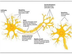
Most common neuron morphology consists of the cell body (the soma), the dendrites that receive messages from other cells, the myelin-covered axon that transmits signals to the axon terminals that communicate with muscles and other nerve cells.
The most common type of motor neuron consists of a cell body, or soma, and an axon that terminates in a series of axon terminals (see figure 1). The soma is the main processing center of the neuron. It is surrounded by dendrites, thin filaments that enable the soma to receive signals from other neurons. The soma processes that information and generates an electrical signal in response. The axon is a long, thin structure that transmits those electrical signals from the soma to another destination in the body, such as sensory organs, muscle fibers, or other neurons. Depending on the function and location, axons can range in length from millimeters to more than a meter. At the far end, the axon splits into multiple axon terminals, enabling a single neuron to communicate with multiple other neurons or muscle fibers. [Note: Other neuron morphologies exist but for purposes of this discussion, we will stick with this most basic model.]
Individual neurons are not physically connected to one another. Instead, they communicate across nanoscale gaps (synapses) between the axon terminals of one neuron and the dendrite spines of another. At the axon terminal, the electrical impulse triggers the release of chemical neurotransmitters that travel across the synaptic gap to receptors on the dendrites of the adjacent neuron. Absorption of the neurotransmitter at the receptor causes the receiving neuron to generate a new electrical impulse, and the signal travels on. Similarly, between motor neurons and the muscles they are trying to control, there is a gap known as the neuromuscular junction. Here, too, neurotransmitters travel across the junction to trigger an electrical signal that causes the muscle to contract or relax.
Neuroplasticity
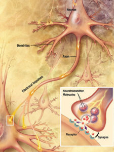
Nerve signals across the synapse via chemical neurotransmitters that travel to receptors on nerve or muscle surface.
Although it’s tempting to think of motor neurons as point-to-point structures, sort of like those tin can phones you might’ve played with as a kid, that’s not exactly the case. Muscles are complex structures built up of individual muscle fibers that are packaged into bundles, which are in turn packaged into larger bundles. Recall that our axons split into multiple terminals. This structure allows a single neuron to excite multiple muscle fibers by using neurotransmitters that bind to receptors on the muscle (see figure 2). In the case of muscles controlling fine motor skills, as with the fingers, a single neuron might only excite a few fibers. In the case of a major muscle group like the gluteus maximus, one neuron might excite hundreds of muscle fibers.
The routes of communication among neurons and between neurons and muscles are not static. Instead, they are constantly changing through neuroplasticity. When neurons are damaged, for example by demyelination, the nervous system uses excess capacity and neuroplasticity to try to find a new path to control the muscle. Intentional movement helps the nervous system establish that pathway, causing the soma to synthesize proteins that enable the dendrites and the axon terminals to grow toward one another. Repetition prompts additional protein synthesis, making the dendrites grow longer and thicker and communicate more effectively. The more frequently one neuron excites a second neuron, the more efficient it gets at exciting that other neuron – cells that fire together wire together, so the saying goes.1,2
This all sounds good so far. The challenge is that neuroplasticity depends on repeating the whole process, from brain signaling to the movement of the body part, and that is not always possible for MS patients. This is where NMES comes into play.
NMES
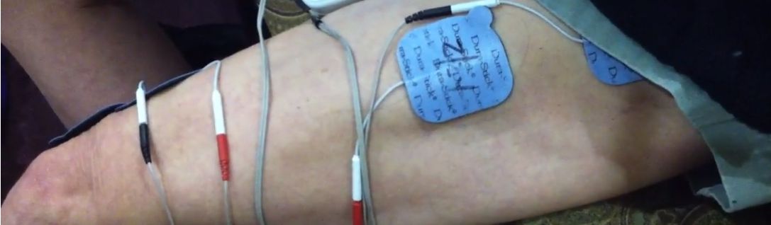
Neurons stimulate the muscles with neurotransmitters that travel from the axon terminal to receptors on the muscle, where they generate electrical impulses that cause the fibers to contract (or relax, depending). NMES uses an external electrical current to substitute for the nerve signal. One electrode goes over the insertion point of the nerve (the neuromuscular junction) and the other goes over the belly of the muscle. This arrangement causes current to flow through the muscle, making it activate. Fingers that normally stay curled can extend, the leg that won’t bend at the knee suddenly flexes. The body wakes up.
If you attempt to move the limb before the current comes on, then run it through full range of motion, NMES can help your nervous system rebuild signaling pathways. It also strengthens muscles through muscle contraction. The result is improved performance as rapidly as the next day from the neural component, followed by increased strength two-to-three days later from the muscular contractions. Performed consistently and strategically, NMES can be used to improve hand function, arm function, balance, posture, sit to stand, walking, and foot drop.
NMES can deliver extraordinary results. The photo sequence below shows the result of a week and a half of work on the hand extensors (see figure 3). The technique requires dedication. Strengthening and building new pathways requires sessions of 45 to 60 minutes. Maintaining function requires sessions of about 15 minutes. The good news is that most of the key muscle groups can be worked while sitting at a desk or on the couch. The other good news is that it feels so good to be able to consciously improve that it doesn’t feel like a hardship.
On a personal note, I discovered NMES in 2013 and I credit it with the fact that after 19 years with PPMS, I can still stand for extended periods of time, I can partially or entirely transfer on my own, I have a moderate amount of hand and arm function, and on a good day I can even scuffle out a few steps. Seldom does a day go by when I don’t work at least a few muscle groups with the technology. If I miss more than two days in a row on a muscle, I can see myself starting to go backward.
There’s no guarantee that the technique will work for everyone but it is worth consideration. Always check with your doctor before considering NMES and always use it under the supervision of a medical professional.
References
- “‘When an axon of cell A is near enough to excite a cell B and repeatedly or persistently takes part in firing it, some growth process or metabolic change takes place in one or both cells such that A’s efficiency, as one of the cells firing B, is increased’” Hebb D. 1949. The organisation of behaviour. New York, NY: John Wiley and Sons. [Google Scholar]
- Shatz CJ. 1992. The developing brain. Sci. Am. 267, 60–67. (10.1038/scientificamerican0992-60)
Disclaimer
Hack My MS provides news and information only. It is an account of my own experiences and some techniques that have worked for me. It should not be construed as medical advice, nor is there any guarantee that any of these techniques will work for you. Always check with a medical professional before starting any exercise program, treatment, or medication. Do not discontinue any exercise program/medication/treatment or delay seeing a doctor as a result of anything you read on this site.
0 Comments